Unveiling Hair Secrets: Microscopy & Analysis [Expert Guide]
Is the seemingly simple strand of hair on your head a silent storyteller, holding secrets of your health and history? The microscopic world of hair, often overlooked, unveils a wealth of information, from the active growth phase to potential health concerns and even forensic clues.
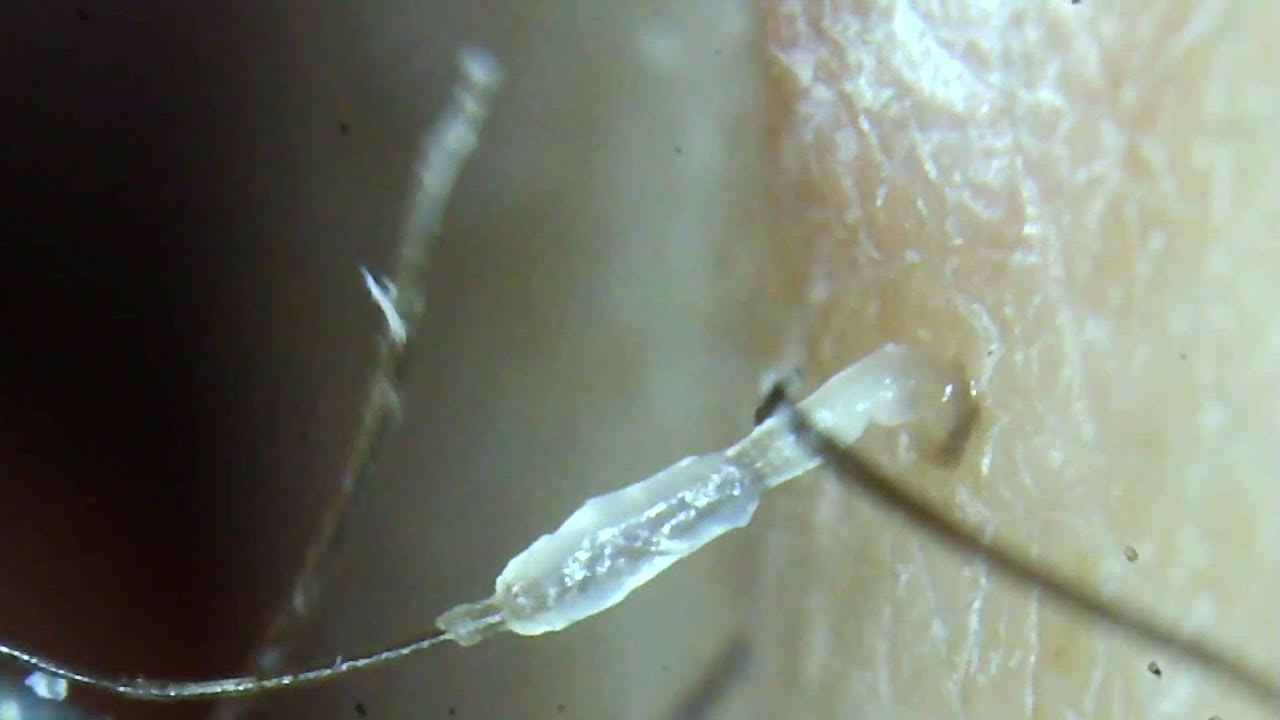
The journey into hair microscopy begins with the seemingly simple act of plucking a hair. When you observe a hair sheath attached to the hair, you've captured a hair in its active growing phase. This is merely the starting point. Equipped with a microscope, microscope slides, and cover slips, the analysis can commence. Whether the hair is attached to its follicle or separated, it can be prepared for examination. If the hair is attached to the follicle, then center it on the slide, if it is not, you can prepare a dry mount of any part of the hair. Applying a few drops of mineral oil to the glass microscope slide can help. This preparation is key to bringing the inner structure of a hair into clear focus.
The applications of hair microscopy are varied and significant. In the realm of dermatology, a trichogram, which is the evaluation of hairs using a microscope, can be a valuable diagnostic tool. This technique allows practitioners to examine hairs for the presence of parasites, like Demodex spp, or louse nits. It is employed for examining hair with obvious defects, which aid in improving our understanding. This may include hair shaft abnormalities caused by disorders or genodermatoses. Changes in the cortex of the hair, for example, can be indicative of underlying health issues. The ability to scrutinize individual hairs under high power offers a window into the overall health and any underlying health issues. For example, a trichogram can help the practitioner examine hairs for parasites (e.g., demodex spp [figure 1]) that may be adjacent to the root of the hair or attached to the hair itself (e.g., louse nits [mallophaga, anoplura]).
In forensic science, the analysis of hair can provide crucial clues. The study of hair under a microscope has real-world applications, extending beyond the classroom setting and into forensic investigations. By scrutinizing the characteristics of hair, such as its color, texture, and structural features, forensic scientists can establish a link between a suspect and a crime scene, or at least, narrow down the pool of possible suspects. The microscopic features of hair offer a unique forensic signature.
The study of hair also extends into research. Hair follicle outer root sheath (ORS) is a putative source of stem cells with therapeutic capacity. ORS contains several multipotent stem cell populations, primarily in the distal compartment of the bulge region. To rule out any adverse effects on the follicles because of the banking process, a sample of plucked hair follicles were collected and analyzed using immunofluorescence using confocal microscopy. The follicles showed no significant difference in protein marker expression after transport and cryopreservation.
The tools and techniques are essential. Begin by plucking a hair from your head. If your hair is attached to the follicle, center it on the slide. If not, you can prepare a dry mount of any part of the hair. View the slide under high power. The light has to pass through the hair specimen to generate a clear microscopic image. A compound microscope has higher power than stereo microscope to resolve the detailed structures of hair. Human hair under the microscope; Human hair under a stereo microscope at 5x magnification, while microscope world hair under a compound microscope can reveal even finer details. The software that is included with the microscope camera we used allows single snapshots and also extended depth of focus, allowing the visualization of multiple planes of focus within the hair shaft.
Hair microscopy, examining the condition of clipped hair shafts or plucked hair under a light microscope, was able to identify abnormalities in the hair shaft, which could aid in improving our understanding. Furthermore, the examination of plucked human hairs contains proliferating cells within the hair sheath (moll, 1996;). They are therefore potentially attractive as an easily accessible tissue in which to assess, the biological state. Understanding the hair shaft allows for a deeper understanding of human health and external influences on our bodies.
Ultimately, the seemingly simple act of examining a strand of hair with a microscope can provide valuable insights into human health, forensic science, and biological research. The world of hair microscopy is not just about identifying abnormalities; it's about understanding the intricate world that resides within each strand.


